
Study on the mechanism of Hippo-YAP signaling pathway regulate autophagy by Beclin1 in phenotypic transition of vascular smooth muscle cells
-
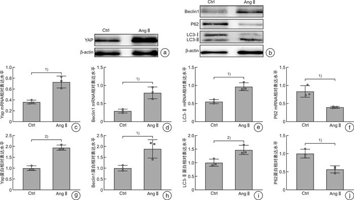
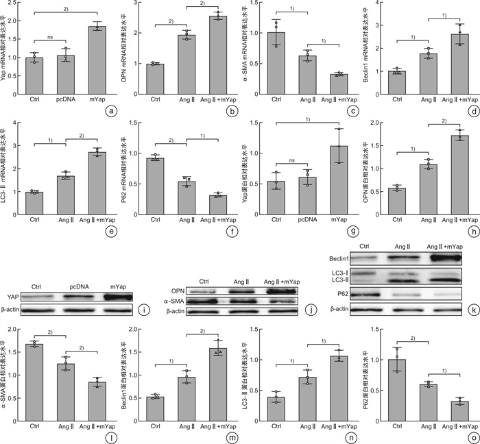
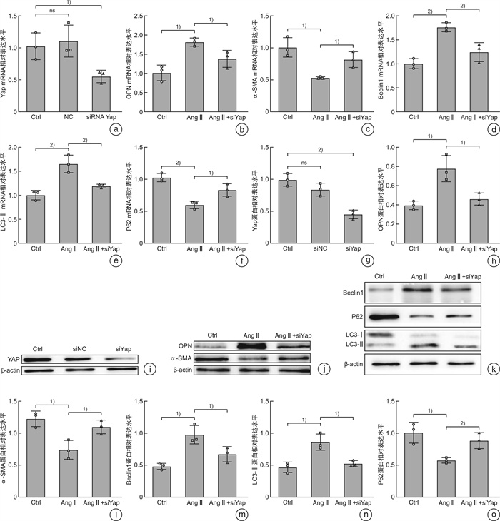
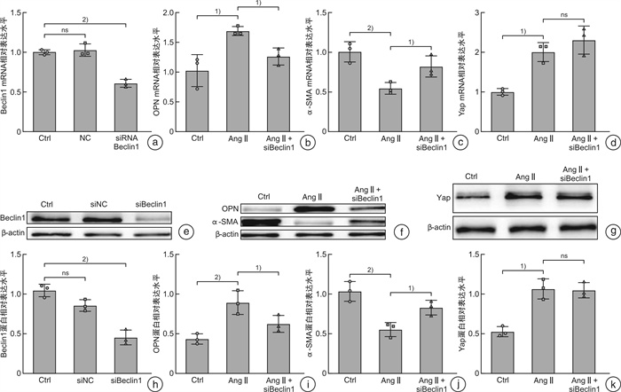
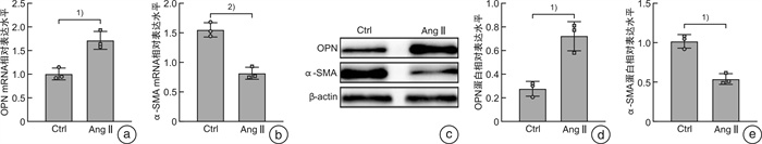
摘要: 目的 探索Hippo-YAP信号通路与自噬在血管平滑肌细胞表型转换中的作用关系及其机制。方法 ① 培养小鼠血管平滑肌细胞(VSMCs),选取第4~8代用于实验,使用血管紧张素Ⅱ(AngⅡ,1×10-7mol/L)处理24 h构建VSMCs表型转换模型,对照组细胞以同浓度的二甲基亚砜(DOSO)处理。②使用Western Blot、qRT-PCR技术在蛋白质及mRNA水平对YAP,VSMCs表型标志物OPN、α-SMA,以及自噬相关标志物LC3、P62、Beclin1进行检测。③分别构建YAP与Beclin1的小干扰RNA(siRNA)对其进行敲减,构建YAP的过表达质粒(pcDNA-YAP)对其进行过表达,使用WB与qRT-PCR验证各基因的表达变化。结果 ① 与对照组相比,AngⅡ处理后VSMCs收缩型标志物α-SMA表达下调、合成型标志物OPN表达上调,YAP及自噬相关标志物LC3Ⅱ、Beclin1表达上调、P62表达下调,在蛋白质及mRNA水平均差异有统计学意义。②与对照组相比,YAP siRNA转染后,VSMCs中YAP在蛋白质与mRNA水平出现明显下调;过表达YAP后,YAP的表达水平出现明显上调。③与单纯AngⅡ刺激组相比,在AngⅡ联合YAP siRNA组中,表型转换的发生及自噬相关标志物的上调明显受到抑制;而过表达YAP组中,表型转换的发生及自噬相关标志物的上调得到进一步增强。④与单纯AngⅡ刺激组相比,AngⅡ联合Beclin1 siRNA组中,表型转换的发生及自噬相关标志物的上调均受到抑制,但两组中YAP的表达变化差异无统计学意义。结论 在VSMCs发生表型转换过程中,自噬相关基因及YAP的表达均明显上调,且YAP可能通过增加Beclin1的表达促进自噬,从而参与表型转换的发生。Abstract: Objective To explore interaction and mechanism between Hippo-YAP signaling pathway and autophagy in phenotypic transition of vascular smooth muscle cells(VSMCs).Methods ① To construct phenotypic transition model of mouse VSMCs, the 4th to 8th generations of VSMCs were treated with angiotensin Ⅱ (AngⅡ, 1×10⁻⁷mol/L) for 24 hours, and the control group was treated with DMSO. ②Western Blot and qRT-PCR were used to detect the expression of YAP, OPN, α-SMA, autophagy-related markers LC3, P62 and Beclin1. ③siRNA was constructed to knock down YAP and Beclin1, respectively. The plasmid was constructed to overexpress YAP. WB and qRT-PCR were used to verify the expression changes of each gene.Results ① Compared to the control group, the expression of α-SMA was significantly down-regulated, While the expression of OPN was up-regulated. YAP and autophagy-related markers LC3Ⅱ、Beclin1 were up-regulated, and the expression of P62 was down-regulated after AngⅡ treatment. There were statistical differences at protein and mRNA levels. ②Compared to the control group, YAP's protein and mRNA in VSMCs were significantly down-regulated after YAP siRNA transfection. After YAP overexpression, YAP was significantly up-regulated. ③Compared to the AngⅡ group, the phenotypic transition and the up-regulation of autophagy-related markers were significantly inhibited in the group treated with both AngⅡ and YAP siRNA. While in the YAP overexpression group, phenotypic transition and up-regulation of autophagy-related markers were further enhanced. ④Compared to the AngⅡ treat group, the phenotypic transition and the up-regulation of autophagy-related markers were inhibited in the group treated with both AngⅡ and Beclin1 siRNA, while the expression of YAP in the two groups was no statistical difference.Conclusion The expression of autophagy-related markers and YAP were up-regulated in VSMCs during phenotypic transitioning, and YAP may promote autophagy by upreglute the expression of Beclin1 and ultimately accelerate phenotypic transition.
-
Key words:
- vascular smooth muscle cells /
- phenotypic transition /
- Yes-associated protein /
- autophagy /
- Beclin1
-

-
表 1 引物序列及产物长度
引物名称 引物序列 产物长度 YAP F:5′-CAAATACAGCTGCAGCAGTTAC-3′ R:5′-CAAATACAGCTGCAGCAGTTAC-3′ 83 bp Beclin1 F:5′-TAATAGCTTCACTCTGATCGGG-3′ R:5′-CAAACAGCGTTTGTAGTTCTGA-3′ 217 bp OPN F:5′-AAACACACAGACTTGAGCATTC-3′ R:5′-TTAGGGTCTAGGACTAGCTTGT-3′ 148bp α-SMA F:5′-CGTGGCTATTCCTTCGTGACTACTG-3′ R:5′-CGTCAGGCAGTTCGTAGCTCTTC-3′ 148 bp LC3 F:5′-CTGTCCTGGATAAGACCAAGTT-3′ R:5′-GTCTTCATCCTTCTCCTGTTCA-3′ 185 bp P62 F:5′-TTCTGGGCCAATCGTTTAAATG-3′ R:5′-ATGCTGCAGAAATACCAACATC-3′ 82 bp GAPDH F:5′-TCAACAGCAACTCCCACTCT-3′ R:5′-TGCTCAGTGTTGGGGGCCGA-3′ 注:YAP:Yes相关蛋白;α-SMA:α-平滑肌肌动蛋白;OPN:骨桥蛋白;LC3:微管相关蛋白1轻链3;GAPDH:三磷酸甘油醛脱氢酶;F:上游引物;R:下游引物。  下载: 导出CSV
-
[1] Hagan PG, Nienaber CA, Isselbacher EM, et al. The International Registry of Acute Aortic Dissection(IRAD): new insights into an old disease[J]. JAMA, 2000, 283(7): 897-903. doi: 10.1001/jama.283.7.897
[2] Yang K, Ren J, Li X, et al. Prevention of aortic dissection and aneurysm via an ALDH2-mediated switch in vascular smooth muscle cell phenotype[J]. Eur Heart J, 2020, 41(26): 2442-2453. doi: 10.1093/eurheartj/ehaa352
[3] Wang F, Chen HZ. Histone Deacetylase SIRT1, Smooth Muscle Cell Function, and Vascular Diseases[J]. Front Pharmacol, 2020, 11: 537519. doi: 10.3389/fphar.2020.537519
[4] Owens GK, Kumar MS, Wamhoff BR. Molecular regulation of vascular smooth muscle cell differentiation in development and disease[J]. Physiol Rev, 2004, 84(3): 767-801. doi: 10.1152/physrev.00041.2003
[5] Chin DD, Poon C, Wang J, et al. miR-145 micelles mitigate atherosclerosis by modulating vascular smooth muscle cell phenotype[J]. Biomaterials, 2021, 273: 120810. doi: 10.1016/j.biomaterials.2021.120810
[6] Ibar C, Irvine KD. Integration of Hippo-YAP Signaling with Metabolism[J]. Dev Cell, 2020, 54(2): 256-267. doi: 10.1016/j.devcel.2020.06.025
[7] 姜文剑, 兰峰, 张宏家. 主动脉血管平滑肌细胞凋亡和Hippo-YAP信号通路作用于主动脉夹层发病的研究进展[J]. 中华胸心血管外科杂志, 2016, 32(1): 51-54. doi: 10.3760/cma.j.issn.1001-4497.2016.01.019
[8] Zhou W, Zhao M. How Hippo Signaling Pathway Modulates Cardiovascular Development and Diseases[J]. J Immunol Res, 2018, 2018: 3696914.
[9] Lin M, Yuan W, Su Z, et al. Yes-associated protein mediates angiotensin Ⅱ-induced vascular smooth muscle cell phenotypic modulation and hypertensive vascular remodelling[J]. Cell Prolif, 2018, 51(6): e12517. doi: 10.1111/cpr.12517
[10] Xie C, Guo Y, Zhu T, et al. Yap1 protein regulates vascular smooth muscle cell phenotypic switch by interaction with myocardin[J]. J Biol Chem, 2012, 287(18): 14598-14605. doi: 10.1074/jbc.M111.329268
[11] Han JH, Park HS, Lee DH, et al. Regulation of autophagy by controlling Erk1/2 and mTOR for platelet-derived growth factor-BB-mediated vascular smooth muscle cell phenotype shift[J]. Life Sci, 2021, 267: 118978. doi: 10.1016/j.lfs.2020.118978
[12] Qi Y, Dai F, Gu J, et al. Biomarkers in VSMC phenotypic modulation and vascular remodeling[J]. Pharmazie, 2019, 74(12): 711-714.
[13] Mondaca-Ruff D, Riquelme JA, Quiroga C, et al. Angiotensin Ⅱ-Regulated Autophagy Is Required for Vascular Smooth Muscle Cell Hypertrophy[J]. Front Pharmacol, 2018, 9: 1553.
[14] Munson MJ, Ganley IG. MTOR, PIK3C3, and autophagy: Signaling the beginning from the end[J]. Autophagy, 2015, 11(12): 2375-2376. doi: 10.1080/15548627.2015.1106668
[15] Kim J, Kundu M, Viollet B, et al. AMPK and mTOR regulate autophagy through direct phosphorylation of Ulk1[J]. Nat Cell Biol, 2011, 13(2): 132-141. doi: 10.1038/ncb2152
[16] Csibi A, Blenis J. Hippo-YAP and mTOR pathways collaborate to regulate organ size[J]. Nat Cell Biol, 2012, 14(12): 1244-1245. doi: 10.1038/ncb2634
[17] Yaghini FA, Song CY, Lavrentyev EN, et al. Angiotensin Ⅱ-induced vascular smooth muscle cell migration and growth are mediated by cytochrome P450 1B1-dependent superoxide generation[J]. Hypertension, 2010, 55(6): 1461-1467. doi: 10.1161/HYPERTENSIONAHA.110.150029
[18] Sherk WM, Khaja MS, Williams DM. Anatomy, Pathology, and Classification of Aortic Dissection[J]. Tech Vasc Interv Radiol, 2021, 24(2): 100746. doi: 10.1016/j.tvir.2021.100746
[19] Milewicz DM, Trybus KM, Guo DC, et al. Altered Smooth Muscle Cell Force Generation as a Driver of Thoracic Aortic Aneurysms and Dissections[J]. Arterioscler Thromb Vasc Biol, 2017, 37(1): 26-34. doi: 10.1161/ATVBAHA.116.303229
[20] Lu QB, Wan MY, Wang PY, et al. Chicoric acid prevents PDGF-BB-induced VSMC dedifferentiation, proliferation and migration by suppressing ROS/NFκB/mTOR/P70S6K signaling cascade[J]. Redox Biol, 2018, 14: 656-668. doi: 10.1016/j.redox.2017.11.012
[21] Zhou C, Lin Z, Cao H, et al. Anxa1 in smooth muscle cells protects against acute aortic dissection[J]. Cardiovasc Res, 2022, 118(6): 1564-1582. doi: 10.1093/cvr/cvab109
[22] Huang B, Niu Y, Chen Z, et al. Integrin α9 is involved in the pathopoiesis of acute aortic dissection via mediating phenotype switch of vascular smooth muscle cell[J]. Biochem Biophys Res Commun, 2020, 533(3): 519-525. doi: 10.1016/j.bbrc.2020.08.095
[23] Liu M, Yu T, Li M, et al. Apoptosis repressor with caspase recruitment domain promotes cell proliferation and phenotypic modulation through 14-3-3ε/YAP signaling in vascular smooth muscle cells[J]. J Mol Cell Cardiol, 2020, 147: 35-48. doi: 10.1016/j.yjmcc.2020.08.003
[24] 陈琳, 程玲霞, 杨帆, 等. 血管平滑肌细胞自噬对小鼠主动脉夹层形成的影响[J]. 中山大学学报(医学科学版), 2021, 42(2): 226-234. https://www.cnki.com.cn/Article/CJFDTOTAL-ZSYK202102010.htm
[25] Clément M, Chappell J, Raffort J, et al. Vascular Smooth Muscle Cell Plasticity and Autophagy in Dissecting Aortic Aneurysms[J]. Arterioscler Thromb Vasc Biol, 2019, 39(6): 1149-1159. doi: 10.1161/ATVBAHA.118.311727
[26] Sun SY, Cao YM, Huo YJ, et al. Nicotinate-curcumin inhibits AngⅡ-induced vascular smooth muscle cell phenotype switching by upregulating Daxx expression[J]. Cell Adh Migr, 2021, 15(1): 116-125. doi: 10.1080/19336918.2021.1909899
[27] Wang N, Xu F, Lu S, et al. Septin4 as an autophagy modulator regulates Angiotensin-Ⅱ mediated VSMCs proliferation and migration[J]. Biochem Biophys Res Commun, 2020, 525(2): 272-279. doi: 10.1016/j.bbrc.2020.02.064
-
图(5)
表(1)
计量
- 文章访问数: 1475
- PDF下载数: 438
- 施引文献: 0