
Magnetization transfer imaging for early assessment of brain injury after cardiopulmonary resuscitation in a rat cardiac arrest model
-
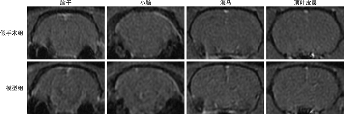
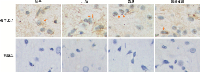
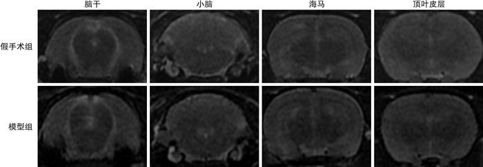
摘要: 目的探讨磁化传递成像(MTI)早期评估窒息大鼠心搏骤停模型复苏(CA/CPR)后脑损伤的可行性。方法将14只大鼠分为假手术组(6只)与模型组(8只),使用气管插管法制作大鼠CA模型,两组大鼠恢复自主循环(ROSC)6 h后行MTI扫描,并测量两组大鼠脑干、小脑、海马及顶叶皮质磁化传递脉冲的信号强度(Ms),并计算磁化传递率(MTR)。MTI扫描完成后将大鼠处死并取脑组织切片行AQP4免疫组织化学染色。比较两组大鼠脑干、小脑、海马及顶叶皮质Ms与MTR差异。结果模型组6只大鼠建模成功,2只失败。模型组大鼠脑干、小脑、海马及顶叶皮质Ms均低于假手术组,两组差异有统计学意义(P < 0.05);模型组测量的各个区域MTR明显高于假手术组,两组差异有统计学意义(P < 0.05)。假手术组大鼠脑干、小脑、海马及顶叶皮质AQP4抗体表达阴性;模型组大鼠脑干、小脑、海马及顶叶皮质AQP4抗体表达阳性。结论MTI技术可早期用于CA/CPR后脑损伤评估,Ms及MTR为敏感反映脑水肿的定量指标。Abstract: ObjectiveTo investigate the feasibility of magnetization transfer imaging(MTI) for early assessment of brain injury after cardiopulmonary resuscitation(CPR) in a rat model of cardiac arrest.MethodsFourteen rats were divided into sham group(n=6) and model group(n=8). The rat CA model was established by tracheal intubation. The two groups of rats underwent MTI 6 hours after recovery of spontaneous circulation(ROSC). Scan and measure the signal strength of magnetization transfer pulses(Ms) in the brainstem, cerebellum, hippocampus and parietal cortex of the rats of two groups, and calculate the magnetization transfer rate(MTR). After the MTI scan, the rats were sacrificed and brain tissue sections were taken for AQP4 immunohistochemical staining. Differences of Ms and MTR in the brainstem, cerebellum, hippocampus and parietal cortex of the two groups of rats were compared.ResultsIn the model group, 6 rats were successfully resuscitated, and 2 rats failed. The Ms in the brainstem, cerebellum, hippocampus and parietal cortex of the model group was lower than that of the sham group, and the MTR of the model group was higher than that of the sham group.The expression of AQP4 in the brainstem, cerebellum, hippocampus and parietal cortex of rats in the sham group was negative; the expression of AQP4 antibody in the brainstem, cerebellum, hippocampus and parietal cortex of rats in the model group was positive.ConclusionMTI technology can be used for early assessment of brain injury after CA/CPR. Ms and MTR are sensitive quantitative indicators reflecting brain edema.
-

-
表 1 两组大鼠基线生理参数
X±S 变量 假手术组 模型组 体重/g 501±13 499±21 心率/(次/ min) 420±12 410±25 平均动脉压/mmHg 119±10 122±14 氧分压/mmHg 92±9 95±11 窒息时间/min - 6±0.8 心脏按压时间/min - 3.9±2.2  下载: 导出CSV
表 2 两组大鼠组内不同脑组织间的MTR值比较
X±S 不同脑区
MTR假手术组 模型组 脑干 0.152075±0.027221 0.224494±0.291766 小脑 0.159930±0.026128 0.205339±0.032136 海马 0.16858±0.154290 0.215533±0.023848 顶叶皮层 0.165003±0.016652 0.208374±0.013355 t 0.541 0.662 P 0.660 0.585
下载: 导出CSV
表 3 两组大鼠Ms比较
X±S 组别 脑干Ms 小脑Ms 海马Ms 顶叶皮质Ms 假手术组 516.31±17.44 506.33±13.72 505.03±10.23 514.83±17.98 模型组 473.68±22.06 485.84±13.64 468.03±16.53 479.26±11.23 t -2.722 -2.593 -4.662 -4.109 P 0.004 0.027 0.001 0.002
下载: 导出CSV
表 4 两组大鼠脑组织MTR比较
X±S 组别 脑干MTR 小脑MTR 海马MTR 顶叶皮质MTR 假手术组 0.152075±0.027221 0.159930±0.026128 0.16858±0.154290 0.165003±0.016652 模型组 0.224494±0.291766 0.205339±0.032136 0.215533±0.023848 0.208374±0.013355 t 4.446 2.686 4.198 4.977 P 0.001 0.023 0.002 0.001
下载: 导出CSV
-
[1] Elmer J, Callaway CW. The Brain after Cardiac Arrest[J]. Semin Neurol, 2017, 37(1): 19-24. doi: 10.1055/s-0036-1597833
[2] Lee BK, Jeung KW, Song KH, et al. Prognostic values of gray matter to white matter ratios on early brain computed tomography in adult comatose patients after out-of-hospital cardiac arrest of cardiac etiology[J]. Resuscitation, 2015, 96: 46-52.
[3] 心肺复苏后昏迷患者早期神经功能预后评估专家共识组. 心肺复苏后昏迷患者早期神经功能预后评估专家共识[J]. 中华急诊医学杂志, 2019, 28(2): 156-162.
[4] Taccone FS, Crippa IA, Creteur J, et al. Estimated cerebral perfusion pressure among post-cardiac arrest survivors[J]. Intensive Care Med, 2018, 44(6): 966-967. doi: 10.1007/s00134-018-5074-3
[5] 刘新峰, 张体江. 磁化传递成像在神经系统疾病的研究现状[J]. 磁共振成像, 2013, 4(2): 146-150. https://www.cnki.com.cn/Article/CJFDTOTAL-CGZC201302022.htm
[6] Dousset V, Grossman RI, Ramer KN, et al. Experimental allergic encephalomyelitis and multiple sclerosis: lesion characterization with magnetization transfer imaging[J]. Radiology, 1992, 182(2): 483-491. doi: 10.1148/radiology.182.2.1732968
[7] 刘志锋, 刘庆余, 叶浩翊, 等. 基于T2WI图像纹理分析评价大鼠心肺复苏后脑损伤初步研究[J]. 中华急诊医学杂志, 2021, 30(12): 1438-1443.
[8] Liu Z, Liu T, Cai J, et al. Quantitative magnetic resonance imaging assessment of brain injury after successful cardiopulmonary resuscitation in a rat model of asphyxia cardiac arrest[J]. Brain Imaging Behav, 2022, 16(1): 270-280. doi: 10.1007/s11682-021-00500-0
[9] 郑阳, 王晓明. 磁化传递成像和酰胺质子转移成像联合评价新生儿脑损伤的初步研究[J]. 磁共振成像, 2017, 8(3): 189-195. https://www.cnki.com.cn/Article/CJFDTOTAL-CGZC201703009.htm
[10] Wang H, Zheng X, Jin J, et al. LncRNA MALAT1 silencing protects against cerebral ischemia-reperfusion injury through miR-145 to regulate AQP4[J]. J Biomed Sci, 2020, 27(1): 40. doi: 10.1186/s12929-020-00635-0
[11] 邵剑波, 沈亚琪, 胡道予, 等. 新生兔脑白质损伤模型磁化传递成像的定量研究[J]. 中华生物医学工程杂志, 2010, 16(5): 402-406.
[12] Shanta SR, Choi CS, Lee JH, et al. Global changes in phospholipids identified by MALDI MS in rats with focal cerebral ischemia[J]. J Lipid Research, 2012, 53(9): 1823-1831.
[13] 韦茂彬, 沈智威, 肖刚, 等. 基于pH值敏感的磁化传递技术在1.5T磁共振成像上的研究[J]. 磁共振成像, 2012, 3(1): 40-43. https://www.cnki.com.cn/Article/CJFDTOTAL-CGZC201201014.htm
[14] Busl KM, Greer DM. Hypoxic-ischemic brain injury: pathophysiology, neuropathology and mechanisms[J]. Neuro Rehabilitation, 2010, 26(1): 5-13.
[15] Tu TW, Lescher JD, Williams RA, et al. Abnormal Injury Response in Spontaneous Mild Ventriculomegaly Wistar Rat Brains: A Pathological Correlation Study of Diffusion Tensor and Magnetization Transfer Imaging in Mild Traumatic Brain Injury[J]. J Neurotrauma, 2017, 34(1): 248-256.
-
图(3)
表(4)
计量
- 文章访问数: 790
- PDF下载数: 171
- 施引文献: 0